Video
First, let’s cover some basic heart anatomy.
Heart anatomy
Atria
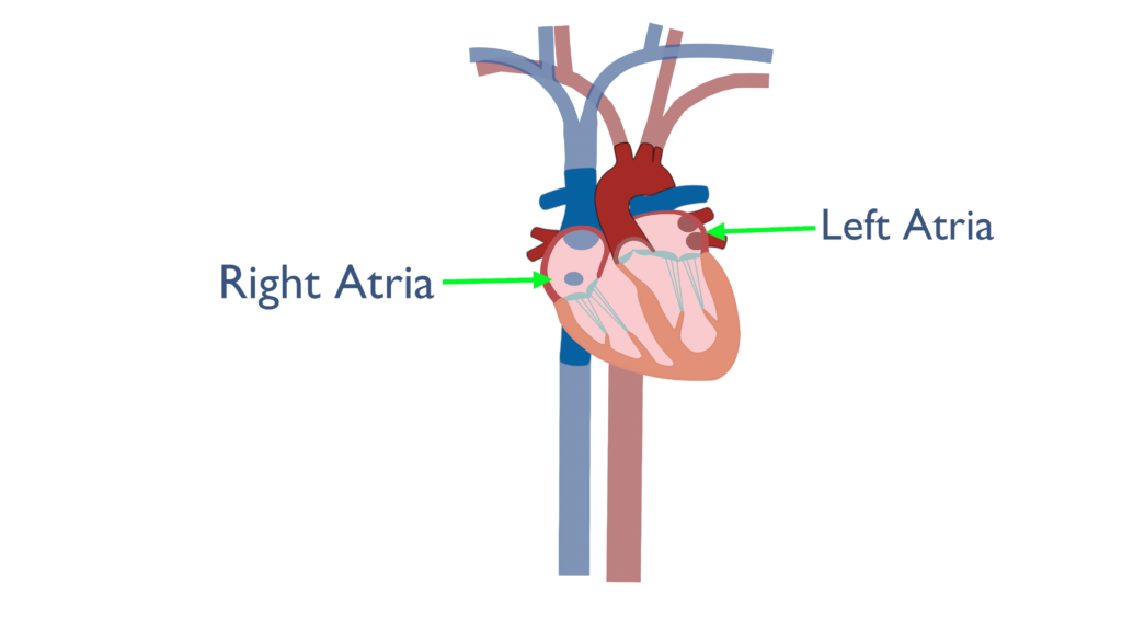
The atria are the top two chambers of the heart. We have a left and right atria just above and connected to the left and right ventricles, respectively.
The purpose of the atria are to force fill the ventricles causing the ventricles to stretch just beyond their maximum resting capacity. This increases contractility of the ventricles and is known as the Frank Starling Law.
Ventricles
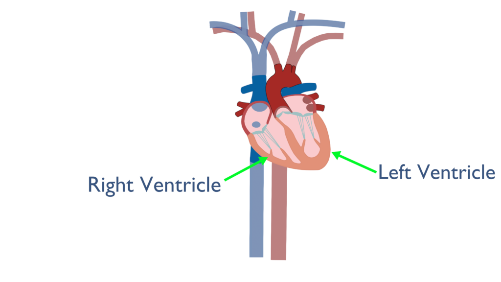
The ventricles are the two bottom chambers of the heart which pump blood throughout the body.
The right ventricle pumps blood to the lungs while the left ventricle pumps blood out to the rest of the body.
The left ventricle is responsible for systemic circulation (blood pumped to the entire body) which is why the left ventricular wall is much thicker than the right ventricle.
Major blood vessels
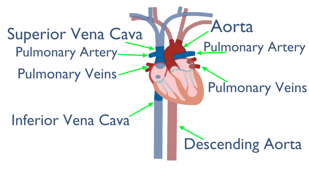
Here we show the most important blood vessels for blood flow throughout the heart and body. Cardiac circulation via the coronary arteries will not be covered here.
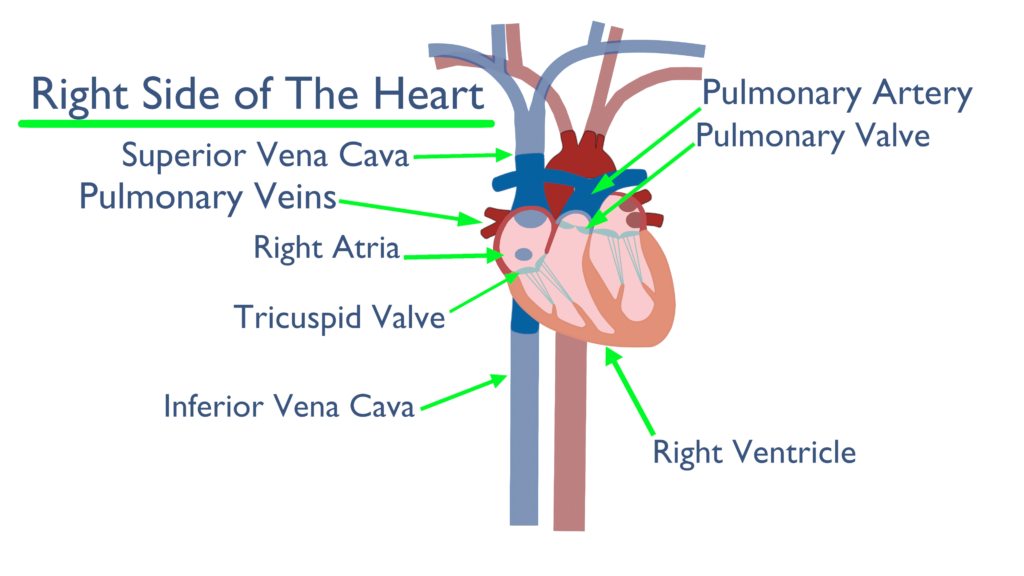
The Vena Cava are responsible to blood return to the heart. The superior and inferior vena cava both connect to the right atria.
The Pulmonary Artery it the vessel which transports blood from the right ventricle to the lungs. This is known as the only de-oxygenated artery within the body. You can see that the Pulmonary Artery branches off to the left and right lungs.
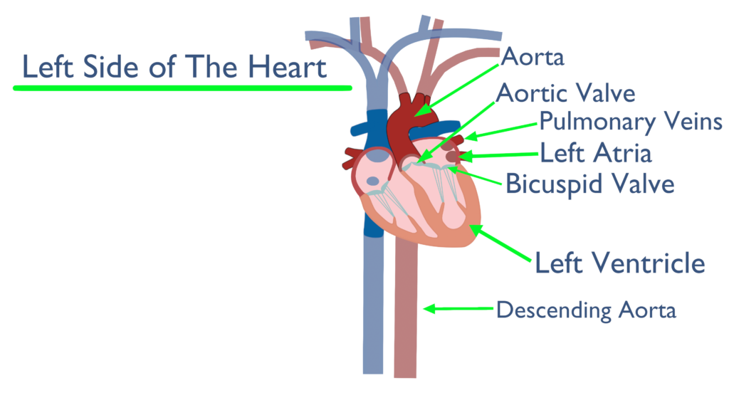
The Pulmonary Veins transport blood from the lungs to the left atria. These are known as the only oxygenated veins within the body. You can see above how they branch into the left atria.
The Aorta is the main artery the transports blood to the entire body. The aortic arch originates from the left ventricle which then wraps above the heart and begins to descend towards the lower body forming the descending aorta. The descending aorta send blood down to the lower body and visceral organs.
Valves
There are four major valves within the heart which are the Aortic Valve, Pulmonary Valve, and the two cuspid valves. These will be covered here.
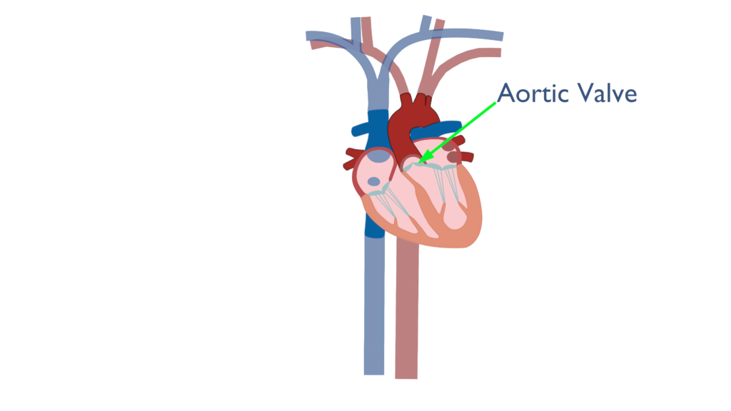
The Aortic Valve separates the left ventricle from the aorta.
When the left ventricle contracts it pushes a bolus of blood through the aortic valve to the aorta, where the blood then pushes through the rest of the body.
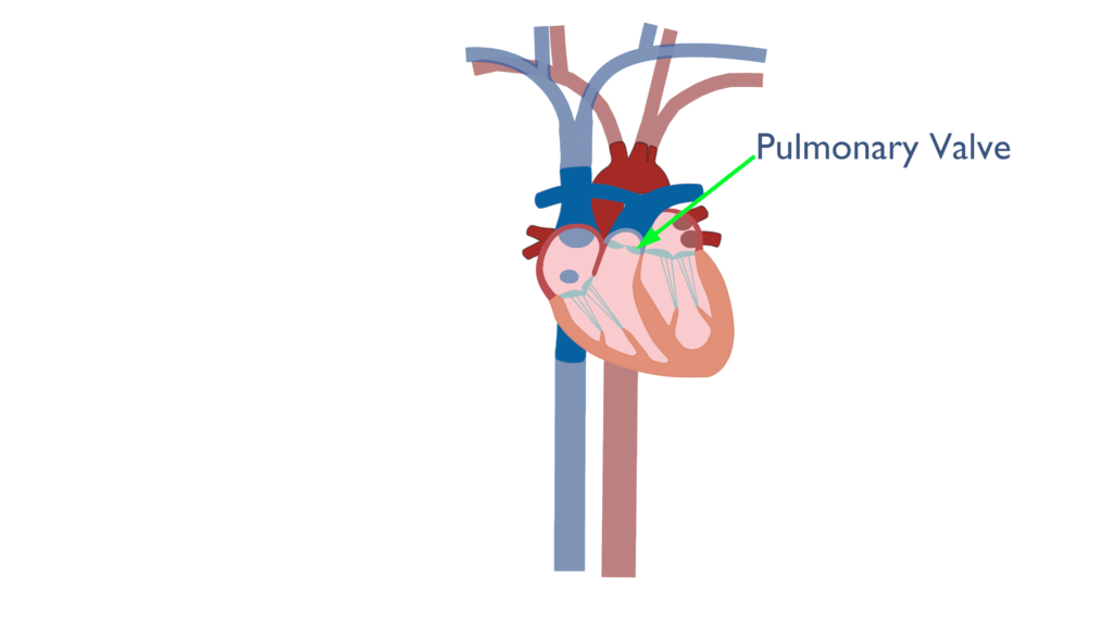
The Pulmonary Valve separates the right ventricle from the pulmonary artery.
When the right ventricle contracts it pushes a bolus of blood to the pulmonary artery, where the blood is then pushed throughout the lungs.
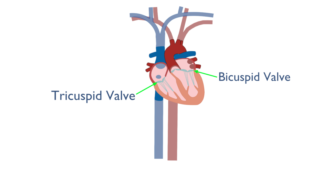
The cuspid valves separate the atria from the ventricles. The cuspid valves are made up of leaflets and small chords (chordae tendinae) that connect the valves to papillary muscles on the ventricles below.
The Tricuspid valve separates the right atria from the right ventricle and is made up of three leaflets, where as, the Bicuspid valve separates the left atria from the left ventricle and is made up of two leaflets.
*The bicuspid valve is also known as the mitral valve.
Direction of blood flow
The way blood flows through the heart is also known as the cardiac cycle. The cardiac cycle is described below:
- Blood enters the atria from either the Vena Cava or the Pulmonary Veins.
- The atria contract forcing the cuspid valves to open and the ventricles to fill with blood.
- The ventricles fill and the cuspid valves close.
- Next there is an isovolumetric contraction phase where the ventricles begin to contract yet the blood volume within the ventricles stays the same.
- This isovolumetric phase is caused due to blood pressure beyond the pulmonary and aortic valves.
- The volume does not change as the ventricles contract until the pressure in the ventricle is greater than the pressure in the arteries beyond the pulmonary and aortic valves.
- Once there is enough pressure within the ventricles the valves will open and blood will begin being ejected from the ventricles.
- The ventricles contract forcing the pulmonary and aortic valves to open.
- Blood is ejected from the ventricles and goes to either the pulmonary artery or the aortic arch.
- The pulmonary and aortic valves close and the ventricles relax.
- Next there is an isovolumetric relaxation phase where the ventricles relax yet the blood volume within the ventricles stays the same.
- During the ventricular isovolumetric relaxation phase the atria begin to refill for the next contraction.