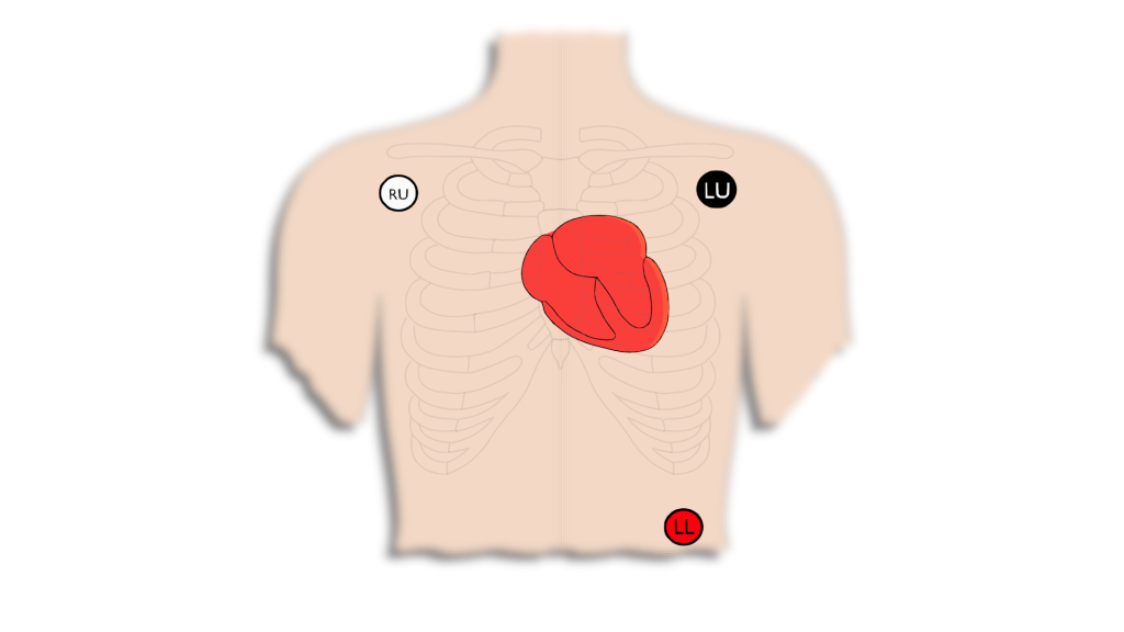
The 3-lead setup is the original system used by Willem Einthoven. This 3-lead concept led to what is known as Einthoven’s Triangle and gives us some really important information regarding the heart.
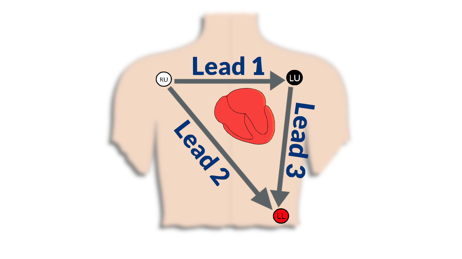
The 3-lead setup consists of the Right Arm/Upper (RU), Left Arm/Upper (LU), and the Left Leg/Lower (LL) leads. From these leads we can see 6 different views (Leads) of the heart. These views are made up of what we know as Einthoven’s triangle (this concept will be very important to understand when evaluating 12-lead ECG).
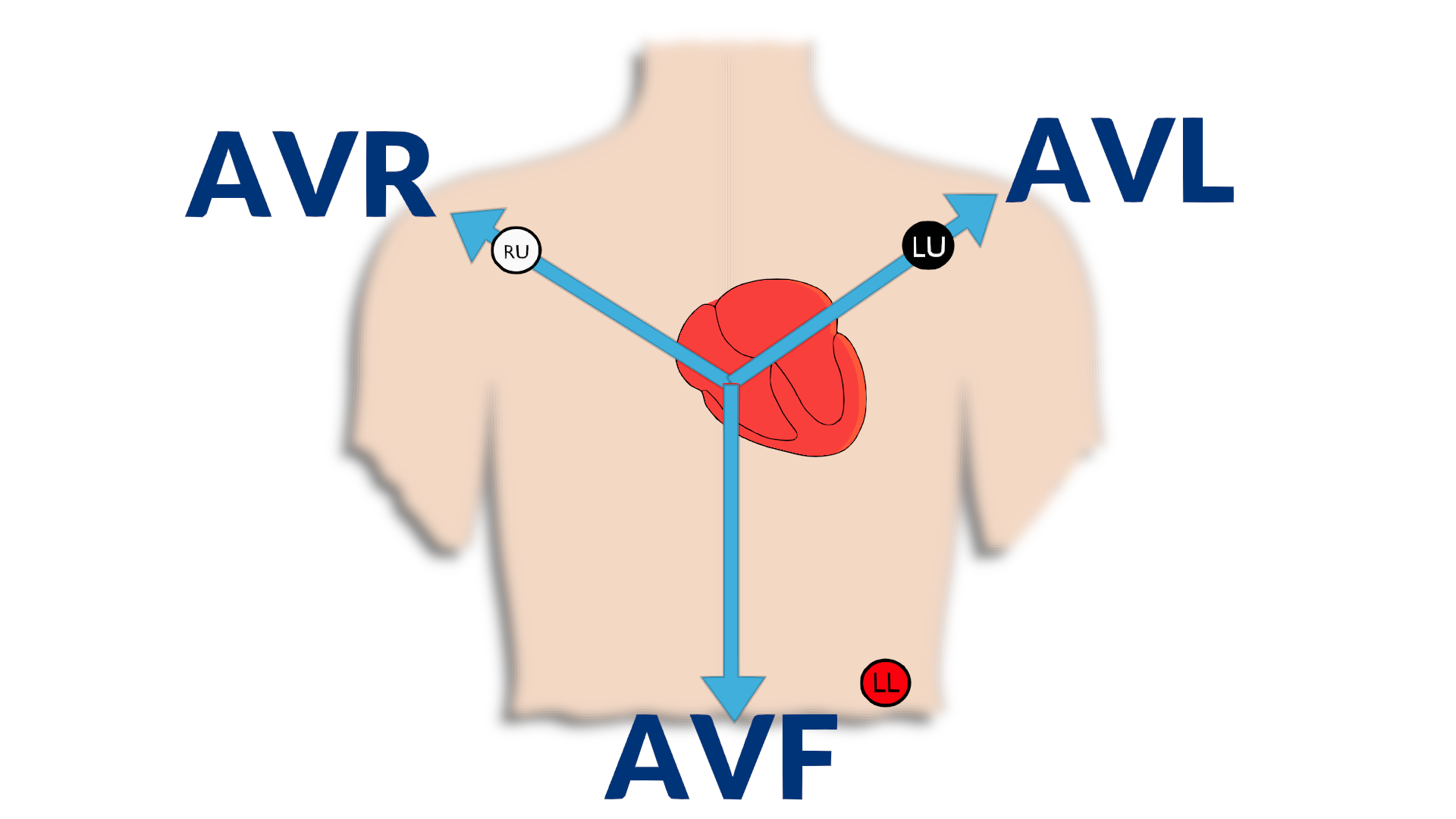
The 6 views we get from Einthoven’s triangle consist of Lead 1, Lead 2, Lead 3, Augmented Vector Right (AVR), Augmented Vector L (AVL), and Augmented Vector Foot (AVF):
- Lead’s 1, 2, and 3 are bipolar in nature, meaning that they are looking at a signal from one lead to another. For example Lead 1 shows the impulse as it travels from the Right Upper lead to the Left Upper lead.
- Augmented Vectors are uni-polar in nature which means they are looking at a view from the heart to a single limb lead.
As we begin to look at rhythm strips we must understand that a positive deflection (i.e. the wave goes upwards) means that the impulse is traveling towards the electrode, whereas if it is a negative deflection (i.e. the wave goes downwards) the impulse is traveling away from the electrode. It is very beneficial to have a good understanding of how the electrical impulse travels through the heart as we begin to talk about the different views.**Click here to refresh on electrical impulse basics**
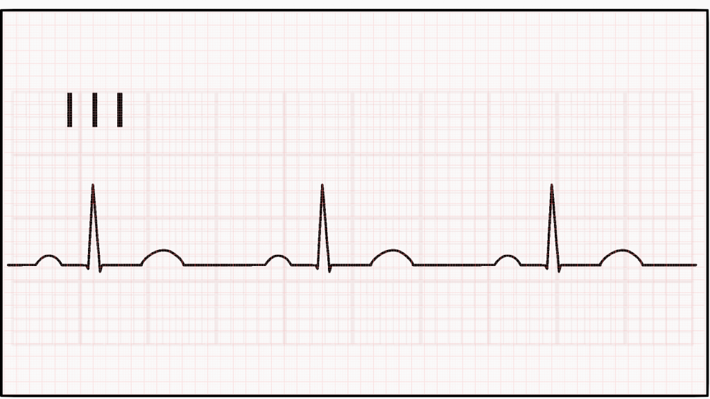
1
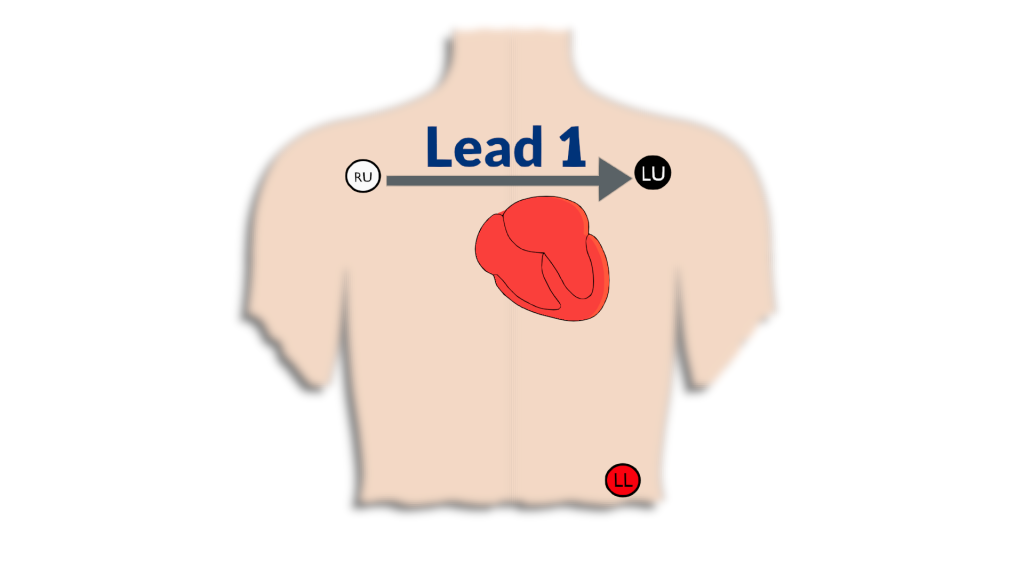
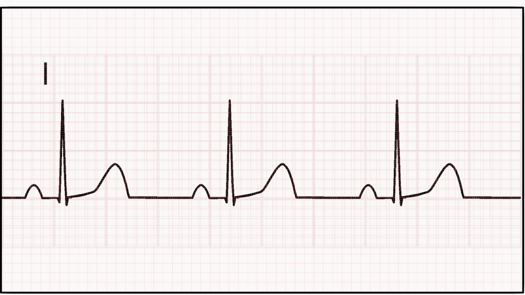
Looking at Lead 1, we see that the P, QRS, and T waves are all positive deflections. This is because we are looking at how the impulse travels from electrode RU to electrode LU which means as the impulse travels from the SA node all the way to the purkinje fibers it is traveling towards LU, so we get all positive deflections.
2
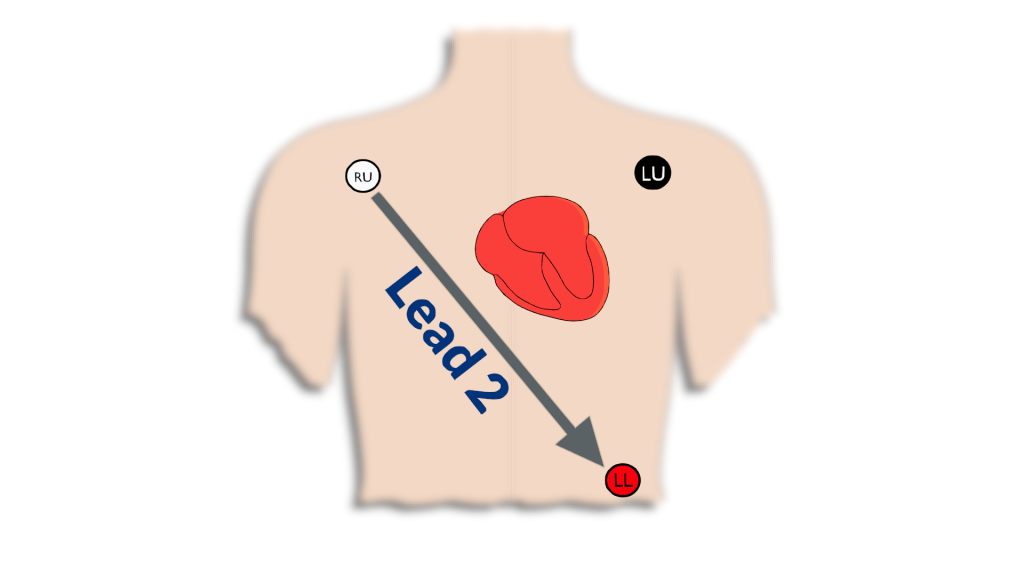
Just as we saw with Lead 1, Lead 2 has all positive deflections since the impulse travels through the heart towards the LL electrode.
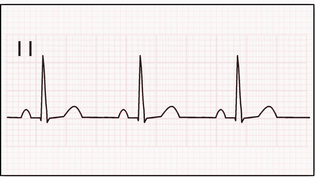
3
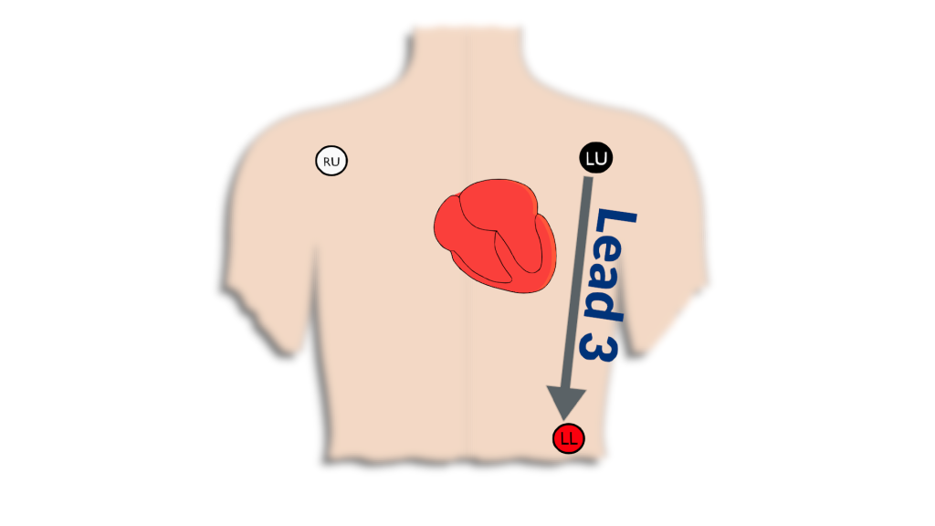
Lead 3 shows the same positive deflections as Leads 1 and 2. Since the view is looking from LU to LL and the electrical impulse is traveling from the top of the heart to the bottom we get a positive deflection within the strip.
AVR
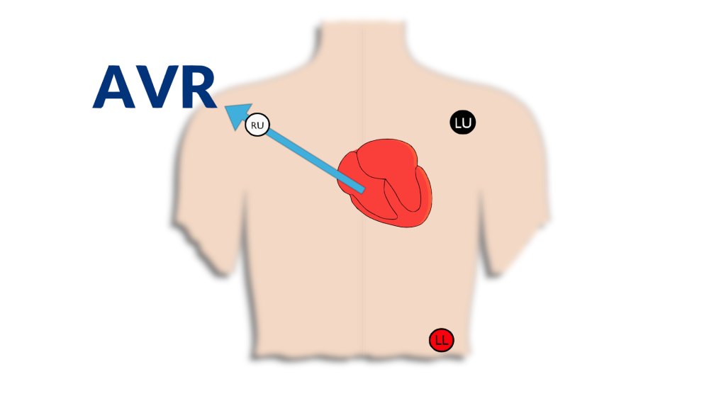
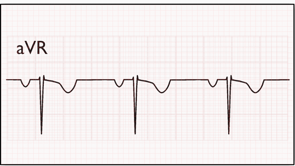
AVR is the first negative deflection we are going to look at. Consider why this may be? This is because AVR is looking towards the RU electrode. Since the impulse travels from the SA node down to the purkinje fibers it is traveling away from the view of the RU electrode, which means the deflections should be negative.
AVL
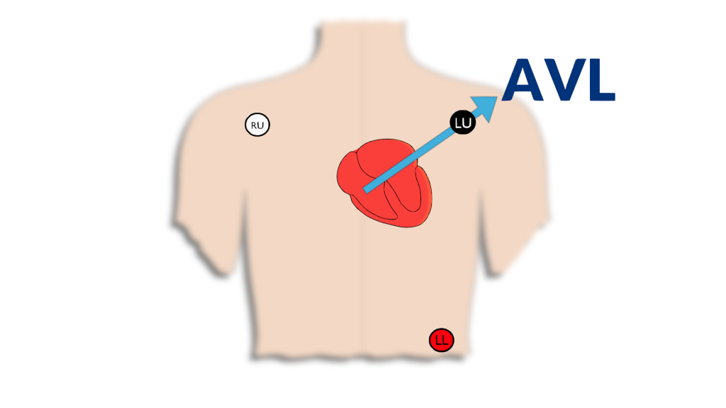
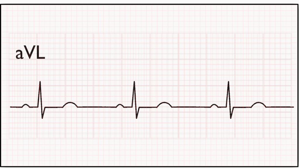
AVL is going to be our first bipolar view (i.e. meaning there should be almost equal positive and negative deflection of the impulse). This is because as the impulse travels from the SA node to the bundle branches it is traveling towards the view, then after passing the bundle branches it begins to travel away from the view. So, we should see positive and negative deflections on the strip.
AVF
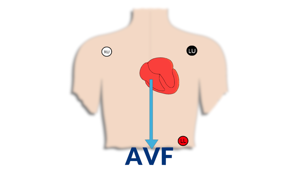
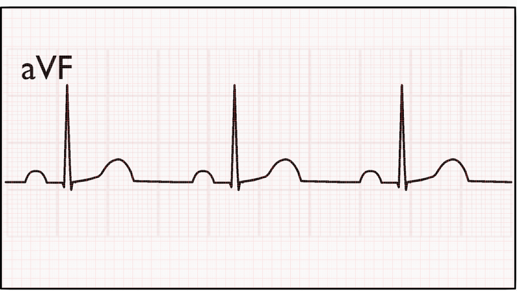
AVF is our full positive augmented view. Since the view is looking straight down the impulse will always be traveling towards the view showing a positive deflection.
Read next: 5 Lead ECG