Video
Below is a quick video snap shot of this article.
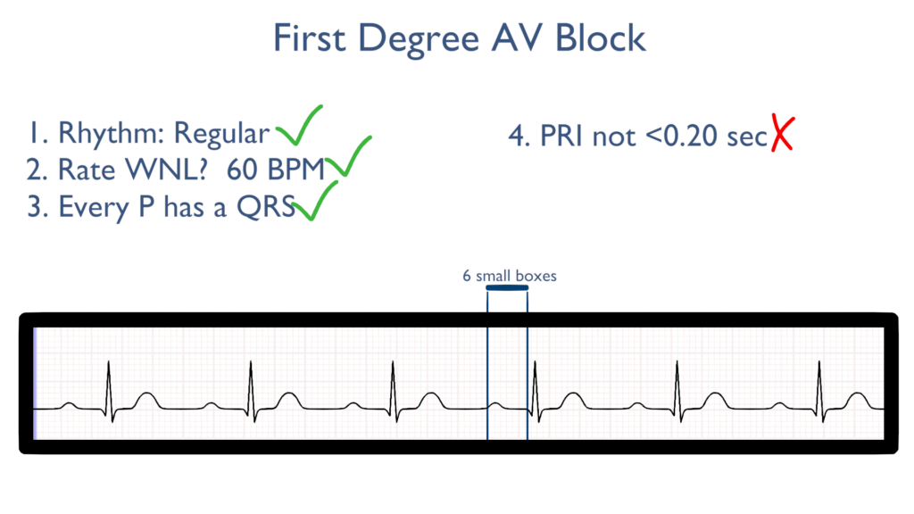
Definition
When there is a prolonged delay in the AV node.
This is easily identified as a PR Interval longer than 0.20 sec or 5 small boxes.
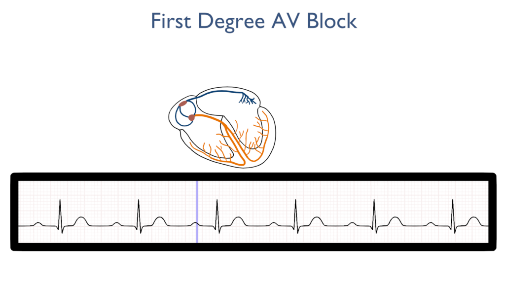
What’s happening in the heart
Atrial Contraction
First, the impulse originates in the SA node just as it does with a normal sinus rhythm.
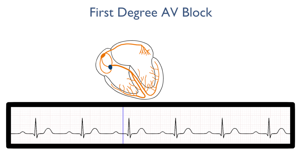
AV Node Delay
Next, the AV node delay happens as expected but lasts longer than expected.
This is identified as a PRI greater than 0.20 sec or 5 small boxes.
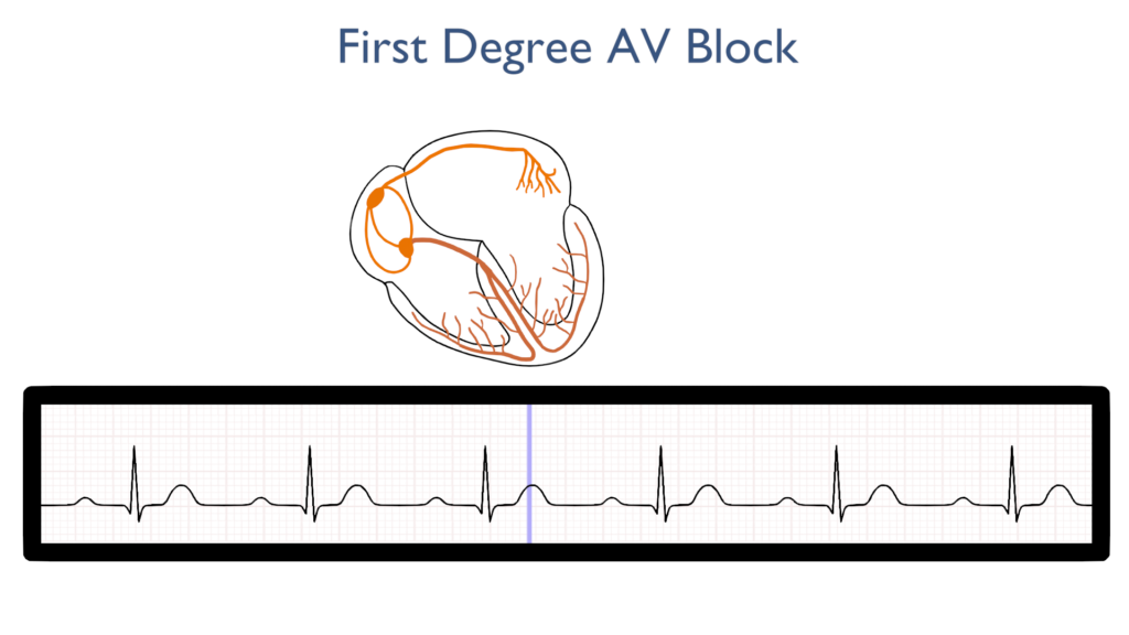
Ventricular Contraction
The QRS complex should be normal with a First Degree AV Block.

Ventricular Repolarization
Lastly, the ventricles relax and reset for the next contraction just as the do with NSR.
T waves should be normal
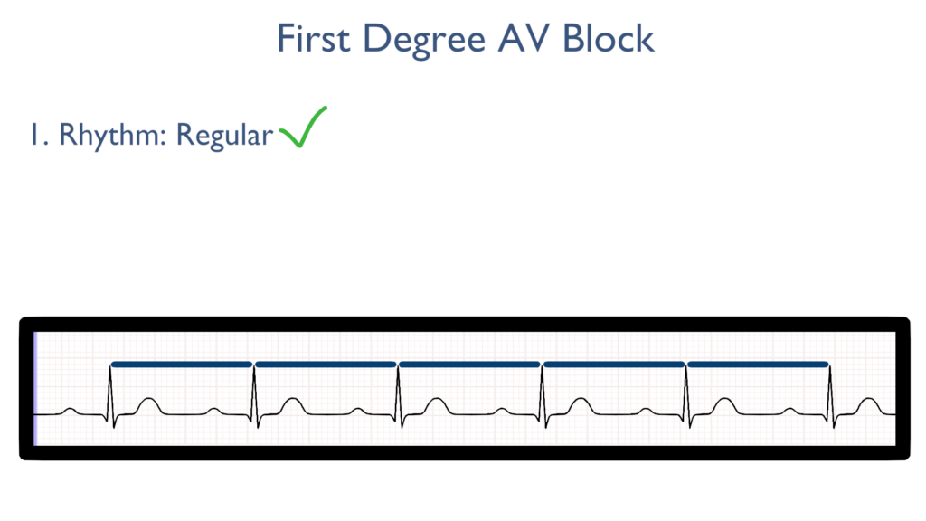
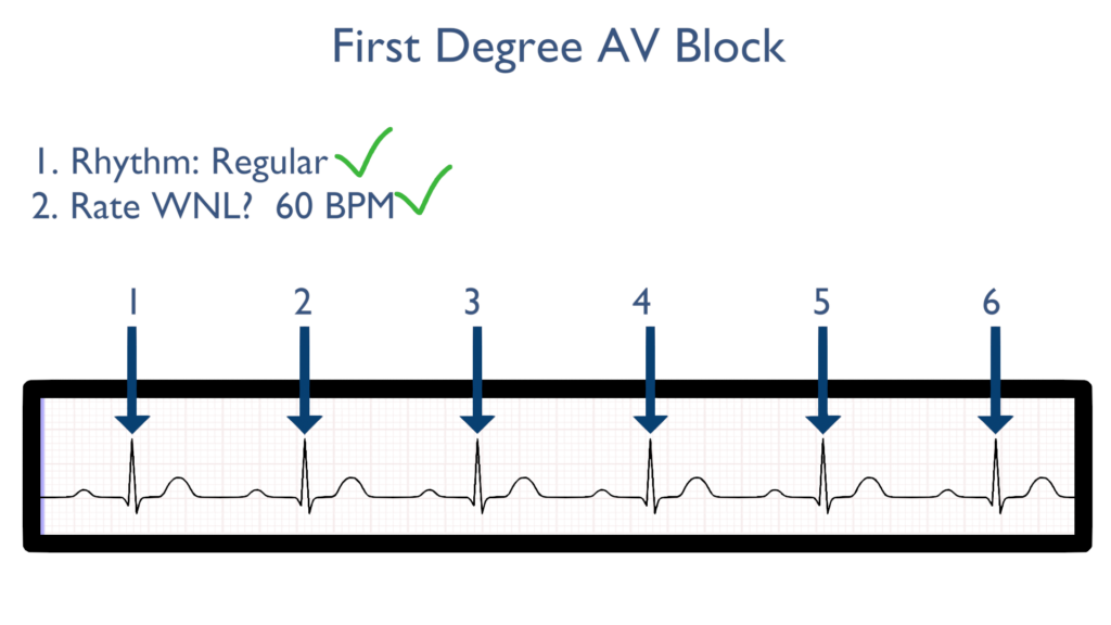
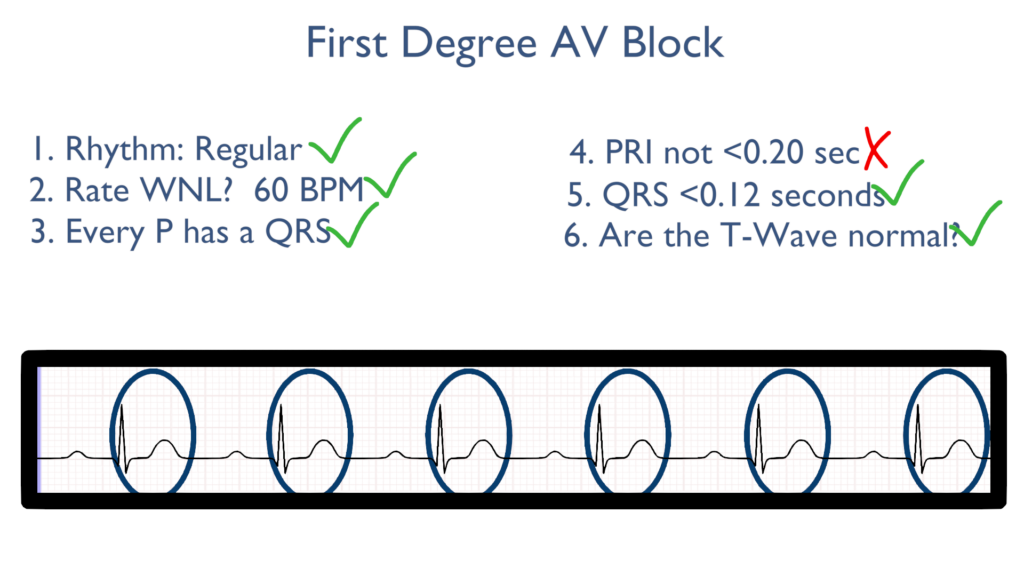
Seven steps of rhythm interpretation
1. Assess the rhythm.
First, the rhythm will be regular.
2. Assess the rate.
Next, when assessing the rate it will most likely be WNL.
3. Assess atrial and ventricular rates.
When it comes to assessing atrial and ventricular rates there will be the same amount of P’s and QRS complexes.
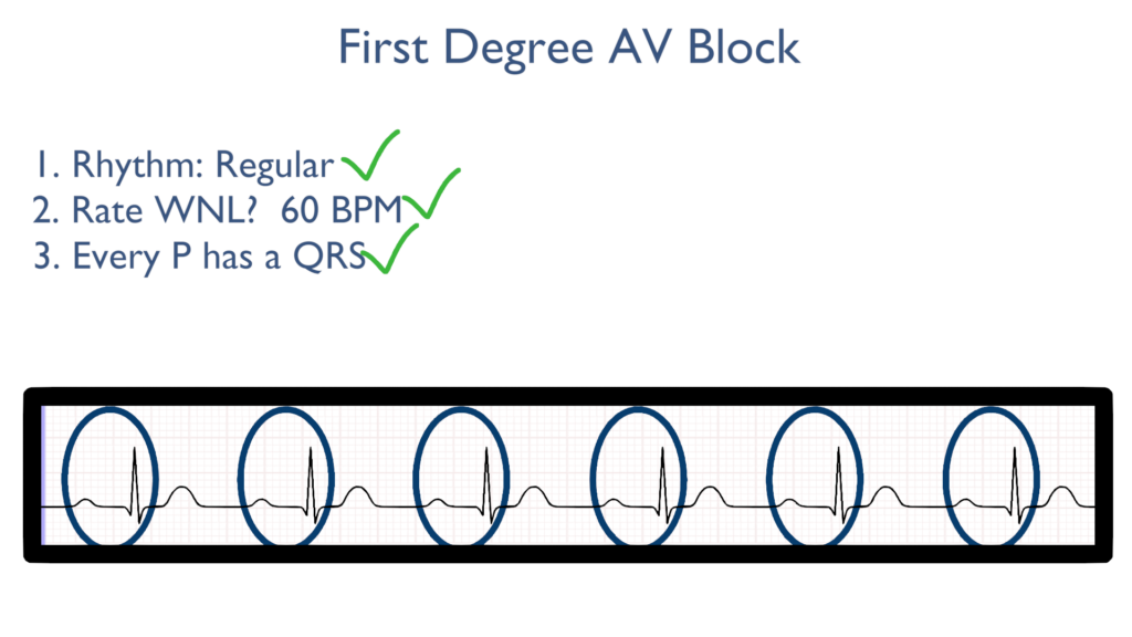
4. Assess the P waves and PR interval.
With a First Degree AV Block there will be a prolonged delay in the AV node causing a greater than normal PR interval.
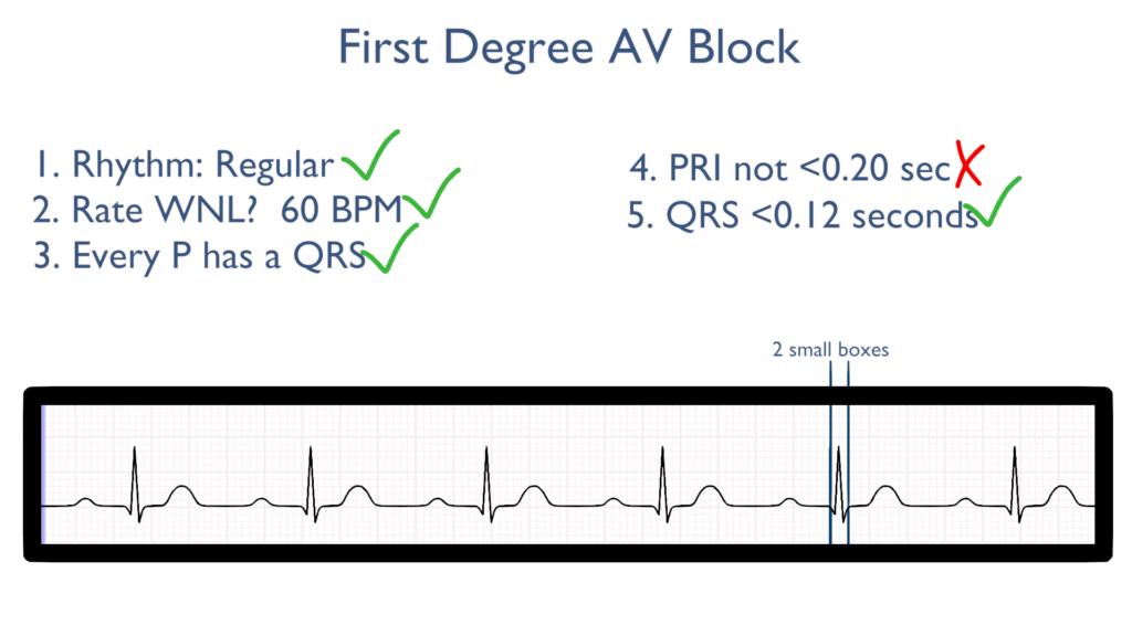
5. Assess the QRS.
Next the QRS will be within normal limits.
The QRS should be between 0.06 seconds to 0.12 seconds.
Better yet, QRS should be between 1.5 to 3 small boxes.
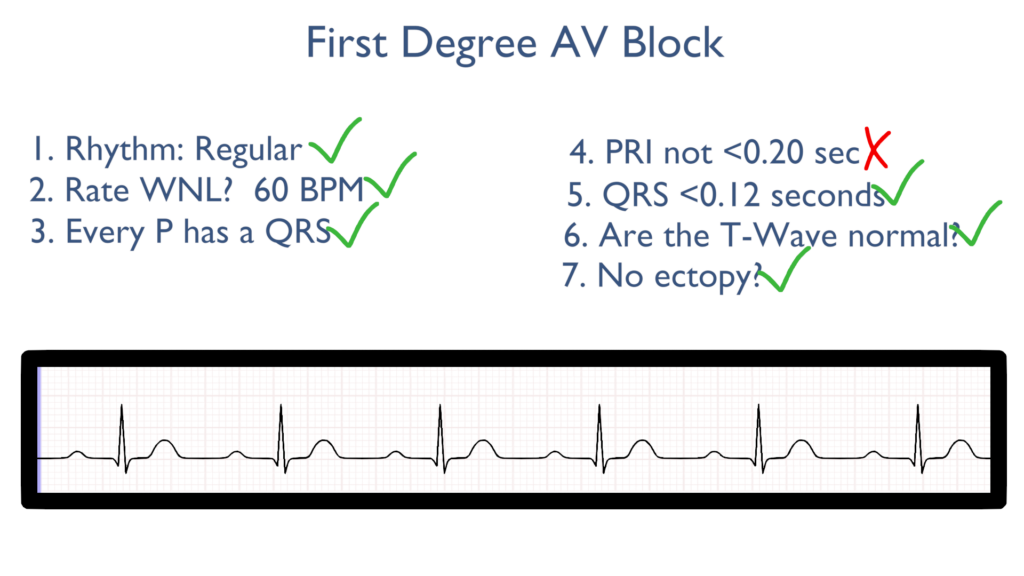
6. Assess the T waves.
T waves should deflect in the positive direction with no ST-Elevation or ST-Depression.
7. Assess for Ectopy.
As you can see on this 6 second strip there are no ectopic beats. This is to say there are no PVC’s or PAC’s present.
Treatments for First Degree AV Block
In most cases first degree AV blocks are benign and do not require treatments, just standard monitoring.