Video
Below is a quick video snap shot of this article.
Definition of Normal Sinus Rhythm
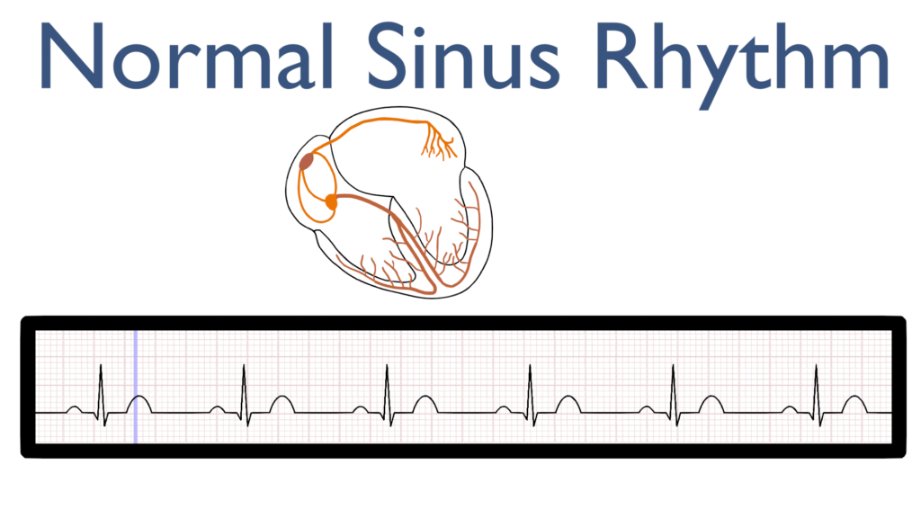
Normal Sinus Rhythm is the typical rhythm that we expect to see. When we talk about normal sinus rhythm we are referring to a rhythm that originates in the SinoAtrial (SA) node and is regular with a rate within normal limits (WNL).
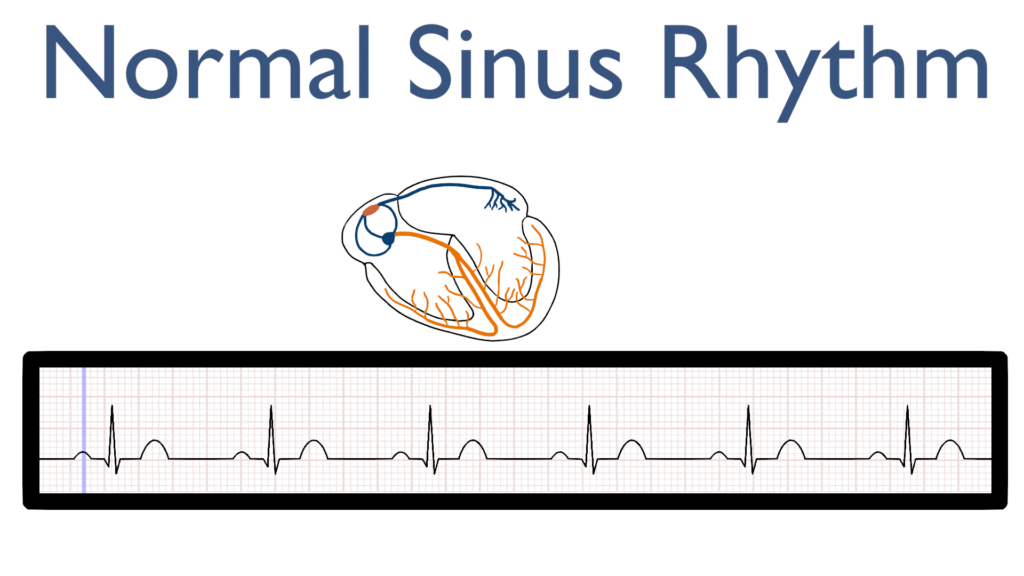
What’s happening in the heart during Normal Sinus Rhythm
Atrial Contraction
First, the impulse originates in the SA node and then travels throughout the Atria.
This is seen as the P-wave on the ECG tracing.
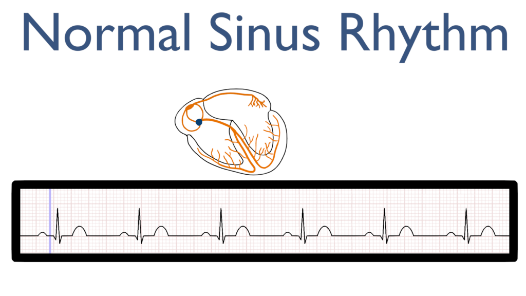
AV Node Delay
Next, there is a delay in the AV node which is seen at the flat line between the P-wave and the QRS complex.
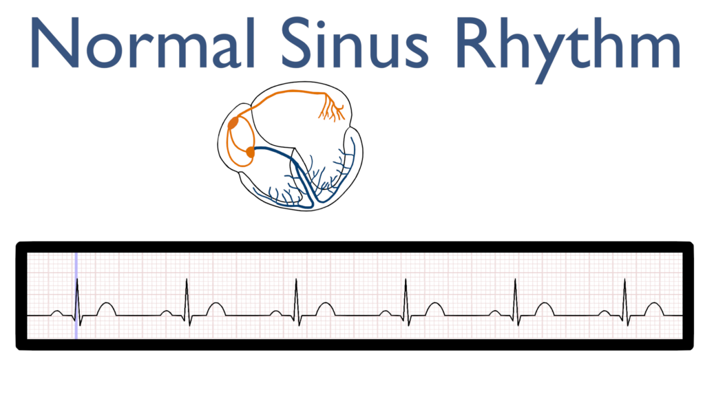
Ventricular Contraction
Next, the impulse travels through the bundle of HIS to the bundle branches and on to the purkinje fibers causing the ventricles to contract.
This is seen as the QRS complex on the ECG tracing.
Ventricular Repolarization
Lastly, the ventricles relax and reset for the next contraction.
This is known as ventricular repolarization and is seen as the T-wave on the ECG tracing.
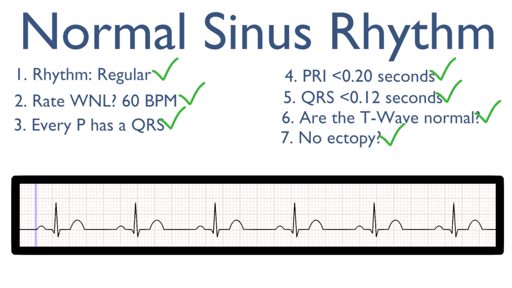
Seven steps of rhythm interpretation for Normal Sinus Rhythm
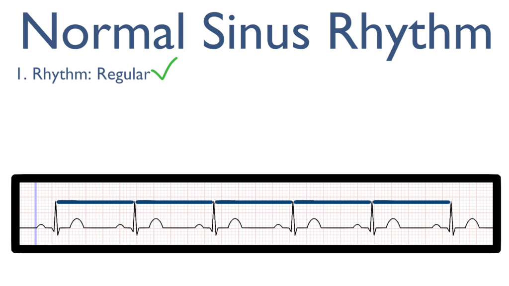
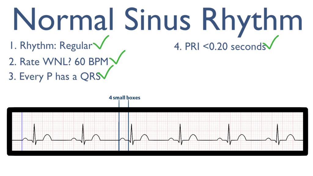
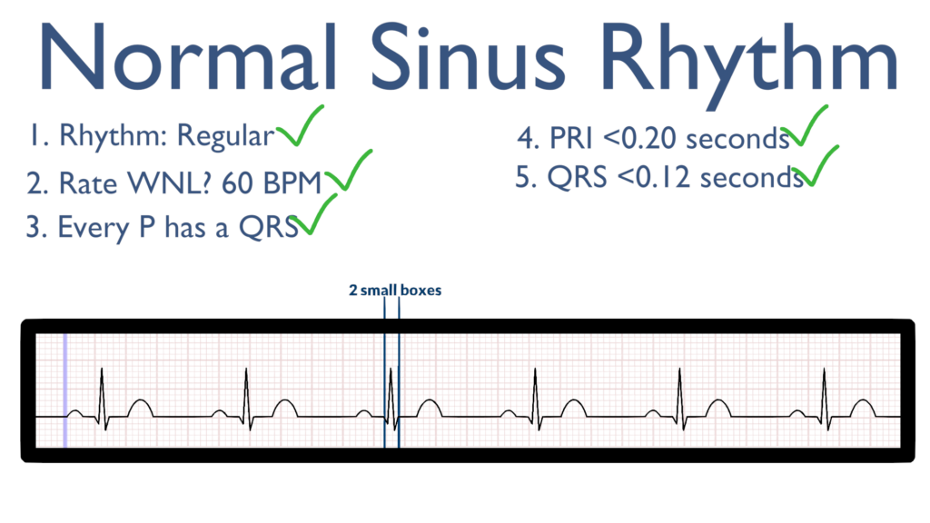
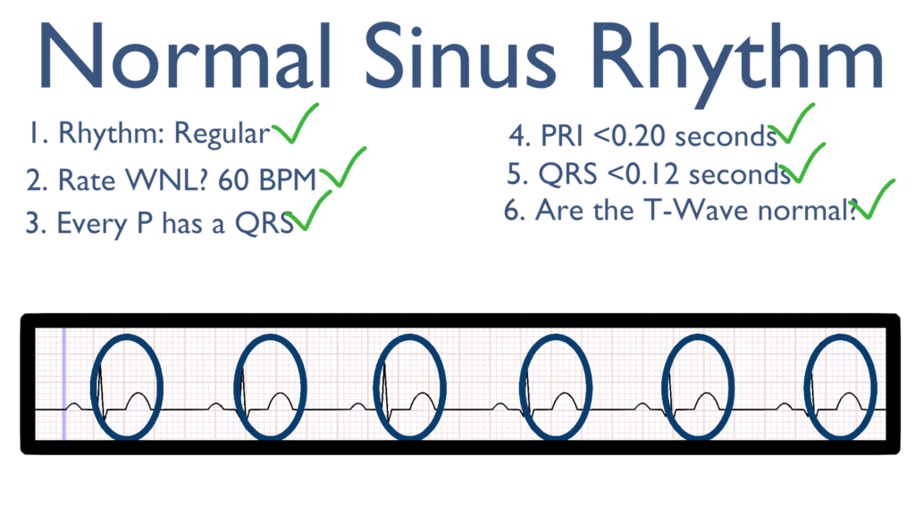
1. Assess the rhythm.
First, with NSR the rate should be regular. This can be identified by regularly spaced R waves.
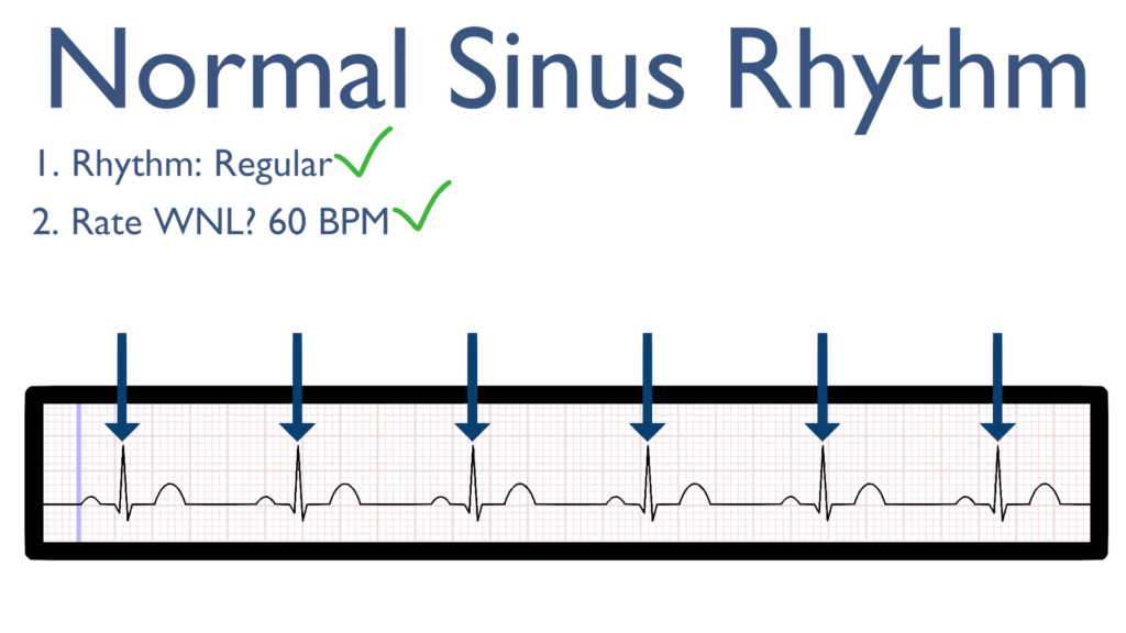
2. Assess the rate.
Next, the rate should be within normal limits (WNL).
We consider a normal heart rate anywhere between 60 to 100 beats per minute (BPM).
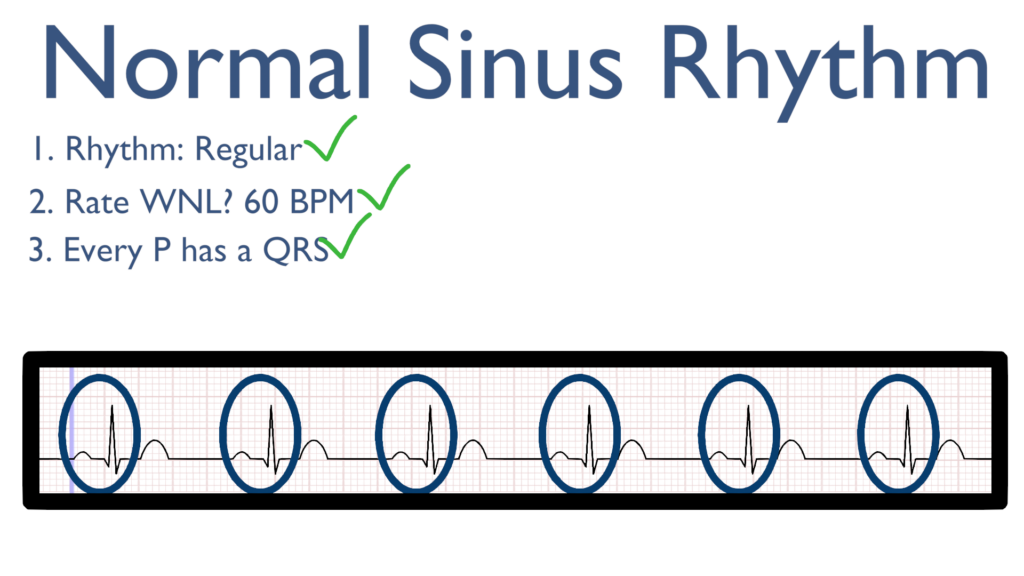
3. Assess atrial and ventricular rates.
Now it’s time to make sure that each QRS has a P, and vice versa, every P should have a QRS (this will make more sense when we evaluate other dysrhythmias),
For NSR we see a QRS directly after every P wave.
4. Assess the P waves and PR interval.
Now take a look at the P waves and assess the PRI. For NSR, all P waves should deflect in the positive direction (upwards).
The PRI should be within 0.12 sec to 0.20 sec.
Better yet, PRI should be between 3 to 5 small boxes.
5. Assess the QRS.
Here take a look at the QRS complex.
The QRS should be between 0.06 seconds to 0.12 seconds.
Better yet, QRS should be between 1.5 to 3 small boxes.
6. Assess the T waves.
For NSR, T waves should deflect in the positive direction with no ST-Elevation or ST-Depression.
The QT Interval should also be between 0.35 seconds to 0.45 seconds.
Easier to remember, the QT interval should be, roughly, between 9 to 12 small boxes.
7. Assess for Ectopy.
As you can see on this 6 second strip there are no ectopic beats. This is to say there are no PVC’s or PAC’s present.
While it would still be considered NSR with ectopy, it is integral to note if there any ectopic beats within the rhythm.