Video
Below is a quick video snap shot of this article.
Definition of Sinus Bradycardia
Sinus Bradycardia (commonly referred to as “brady”) is the same as NSR but the rate is slower than 60 beats per minute (BPM).
What’s happening in the heart
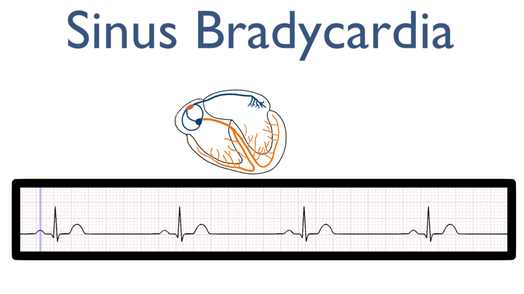
Atrial Contraction
First, the impulse originates in the SA node and then travels throughout the Atria.
This is seen as the P-wave on the ECG tracing.
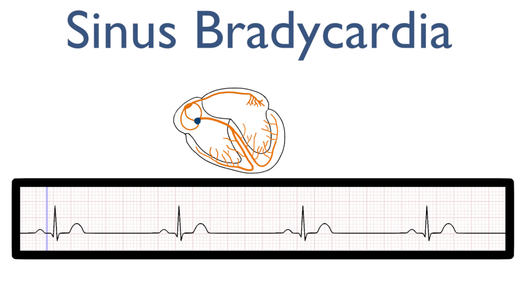
AV Node Delay
Next, there is a delay in the AV node which is seen at the flat line between the P-wave and the QRS complex.
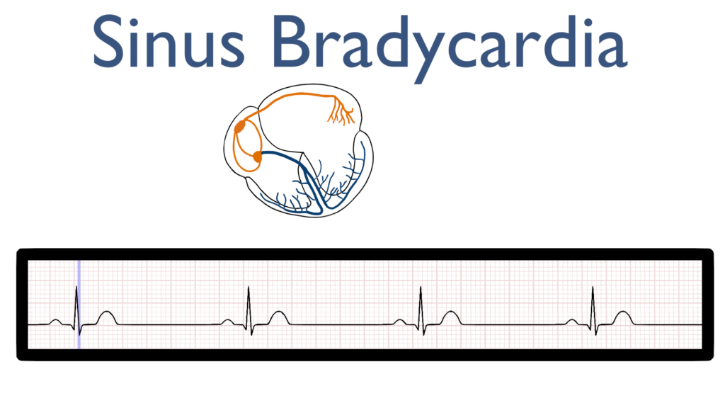
Ventricular Contraction
Next, the impulse travels through the bundle of HIS to the bundle branches and on to the purkinje fibers causing the ventricles to contract.
This is seen as the QRS complex on the ECG tracing.
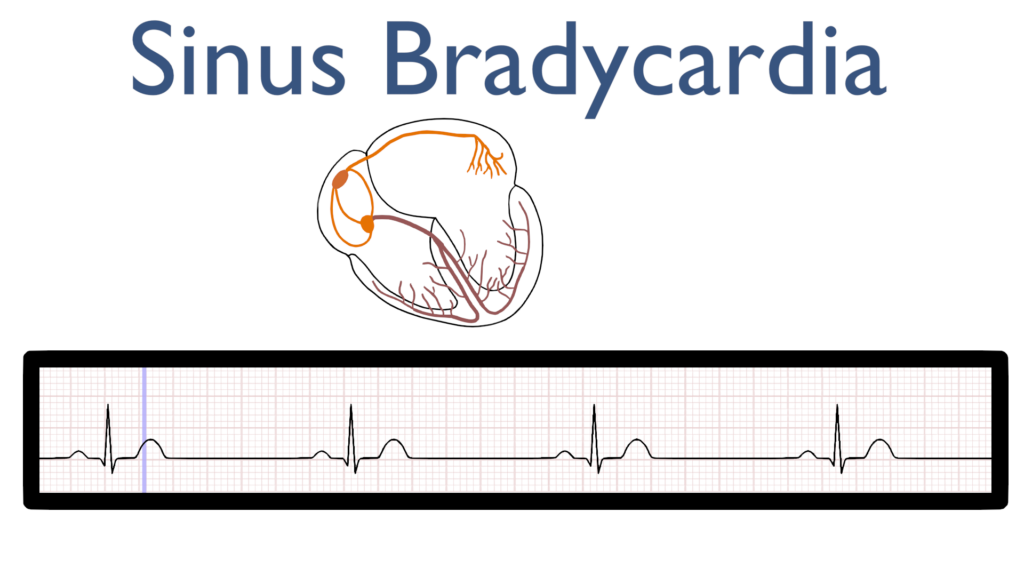
Ventricular Repolarization
Lastly, the ventricles relax and reset for the next contraction.
This is known as ventricular repolarization and is seen as the T-wave on the ECG tracing.
Seven steps of rhythm interpretation
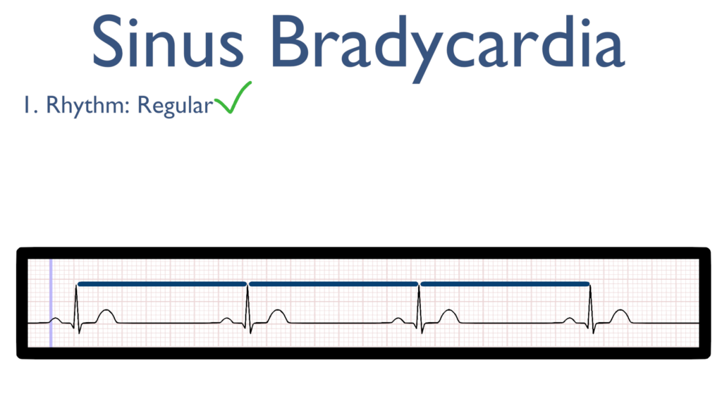
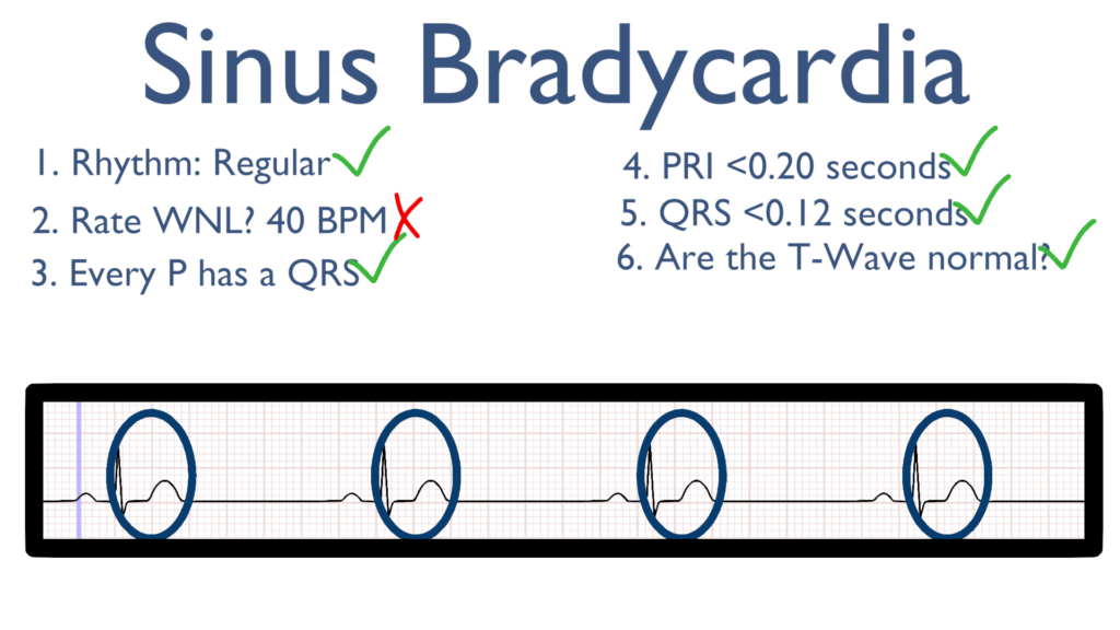
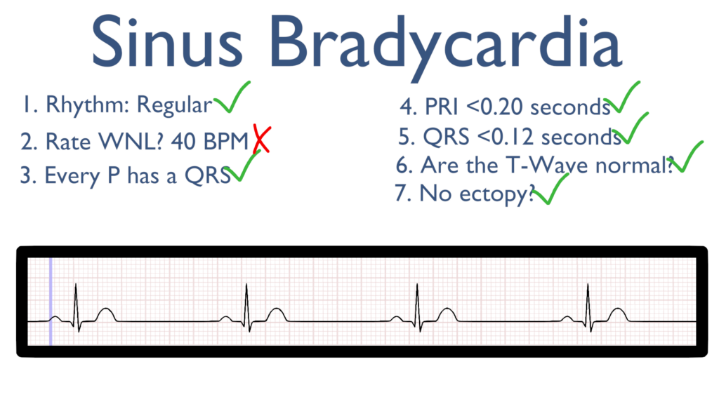
1. Assess the rhythm.
First, the rate should be regular. This can be identified by regularly spaced R waves.
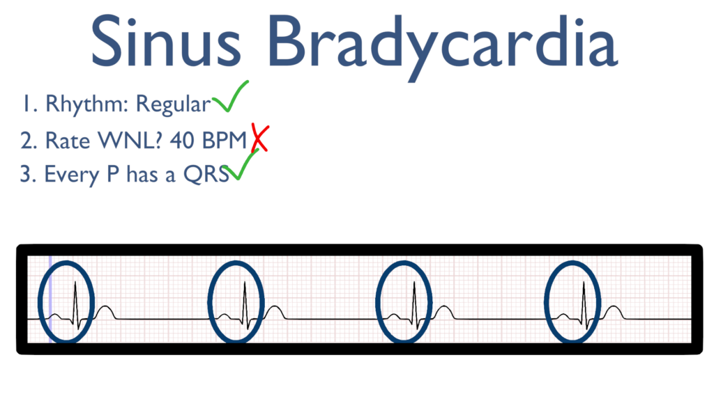
2. Assess the rate.
Next, when assessing the rate you will find it is below 60 bpm.

3. Assess atrial and ventricular rates.
For Sinus Brady we see a QRS directly after every P wave.
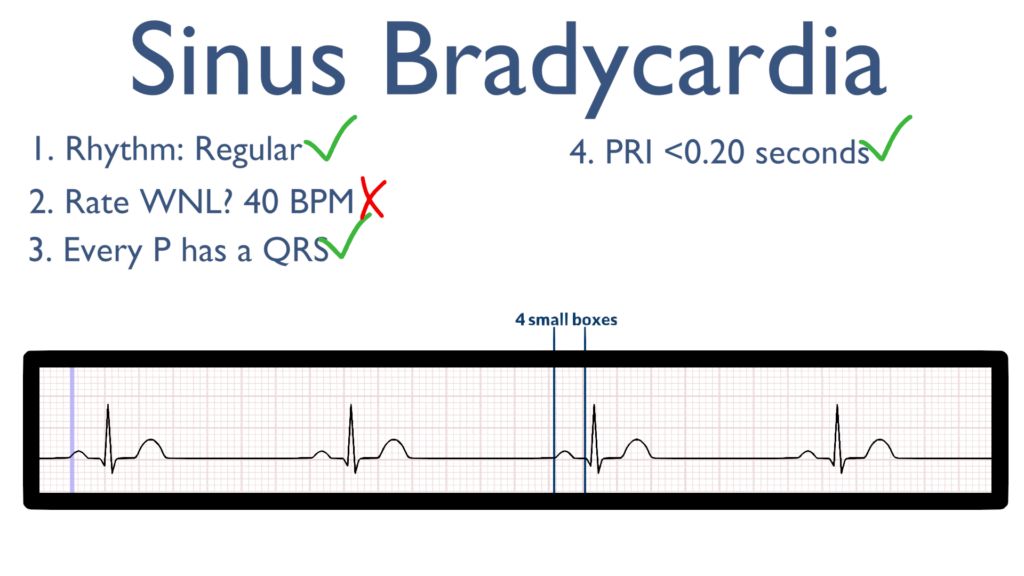
4. Assess the P waves and PR interval.
Now take a look at the P waves and assess the PRI. For Sinus Brady, all P waves should deflect in the positive direction (upwards).
The PRI should be within 0.12 sec to 0.20 sec.
Better yet, PRI should be between 3 to 5 small boxes.
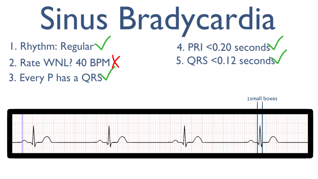
5. Assess the QRS.
Here take a look at the QRS complex.
The QRS should be between 0.06 seconds to 0.12 seconds.
Better yet, QRS should be between 1.5 to 3 small boxes.
6. Assess the T waves.
T waves should deflect in the positive direction with no ST-Elevation or ST-Depression.
The QT Interval should also be between 0.35 seconds to 0.45 seconds.
Easier to remember, the QT interval should be, roughly, between 9 to 12 small boxes.
7. Assess for Ectopy.
As you can see on this 6 second strip there are no ectopic beats. This is to say there are no PVC’s or PAC’s present.
Treatments for Bradycardia
Stable
i.e. asymptomatic and normotensive
Unstable
i.e. hypotensive with or without symptoms
Atropine. 1 mg every 3-5 minutes for a total of 3 doses
Epinephrine drip.
Dopamine drip.
Transcutaneous pacing and pacemaker placement may be needed.