Video
Below is a quick video snap shot of this article.
Definition of Sinus Tachycardia
Sinus Tachycardia (commonly referred to as “tachy”) is the same as NSR but the rate is greater than 100 beats per minute (BPM).
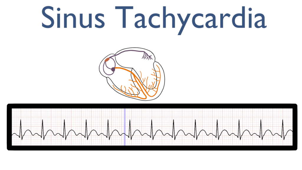
What’s happening in the heart
Atrial Contraction
First, the impulse originates in the SA node and then travels throughout the atria just as it does with NSR
This is seen as the P-wave on the ECG tracing.
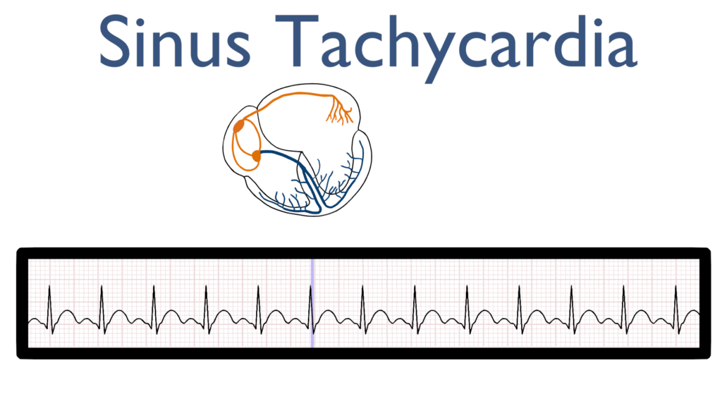
AV Node Delay
Next, there is a delay in the AV node which is seen at the flat line between the P-wave and the QRS complex.
When comparing the AV delay from Sinus Tach to NSR you’ll see that the delay is negligible and appears to not exist since the impulse is traveling so fast.

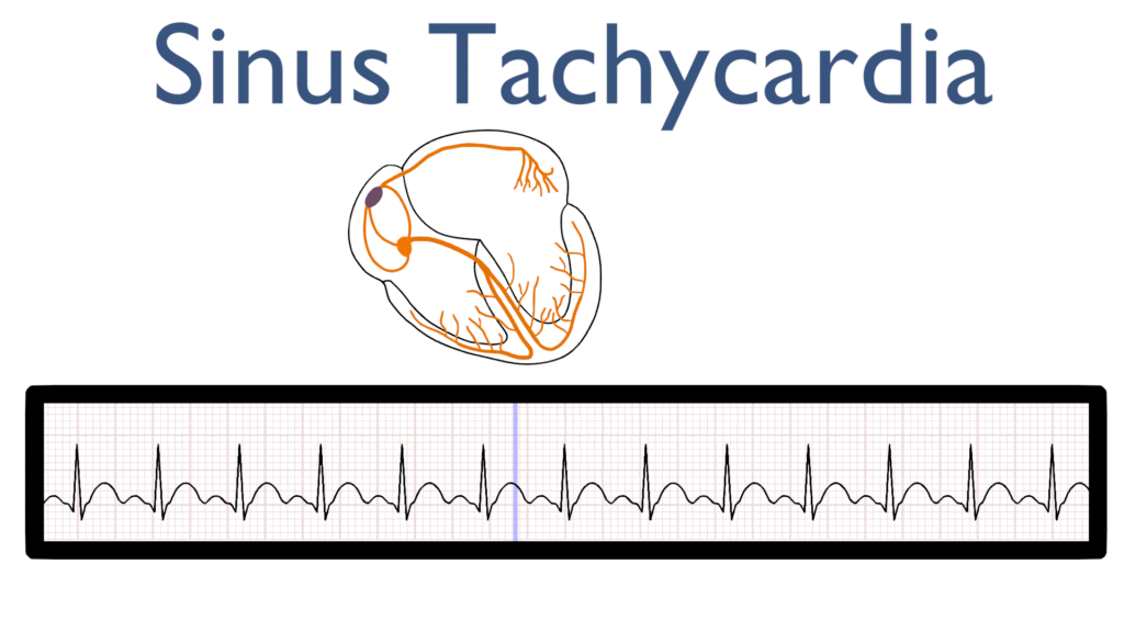
Ventricular Contraction
Next, the impulse travels through the bundle of HIS to the bundle branches and on to the purkinje fibers causing the ventricles to contract.
This is seen as the QRS complex on the ECG tracing.
Ventricular Repolarization
Lastly, the ventricles relax and reset for the next contraction.
This is known as ventricular repolarization and is seen as the T-wave on the ECG tracing.
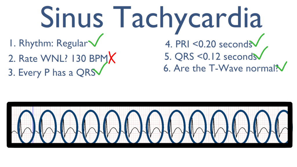
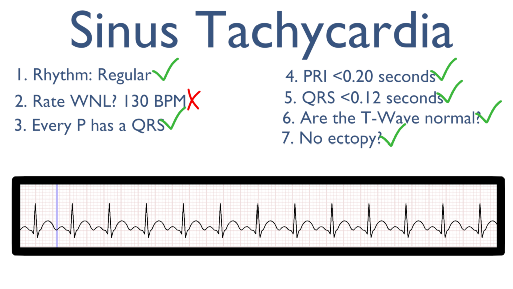
Seven steps of rhythm interpretation
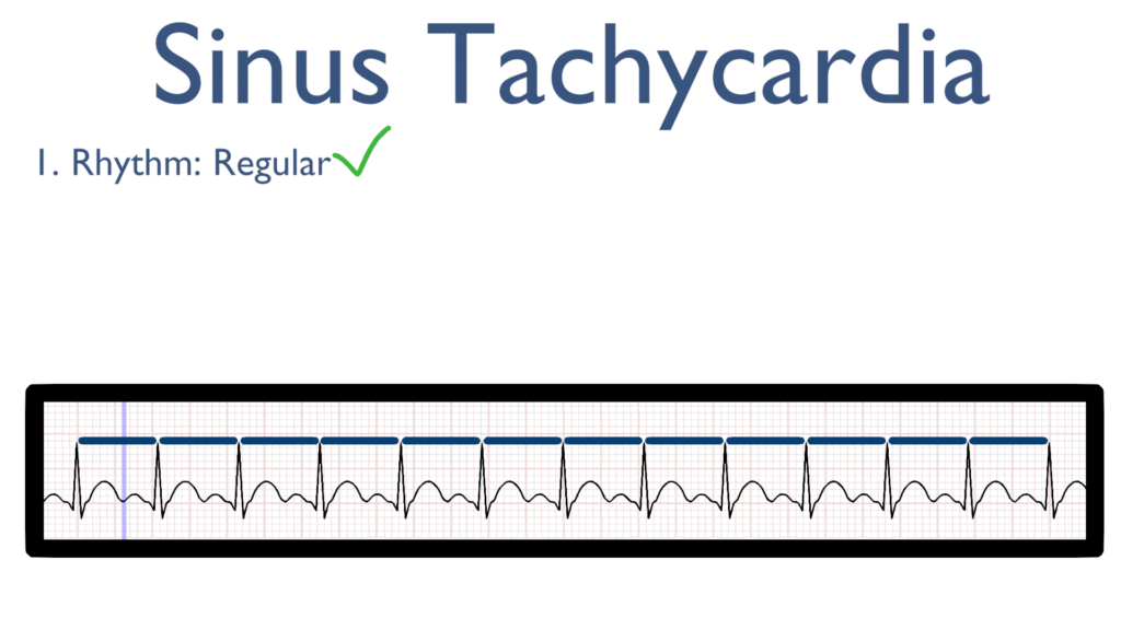
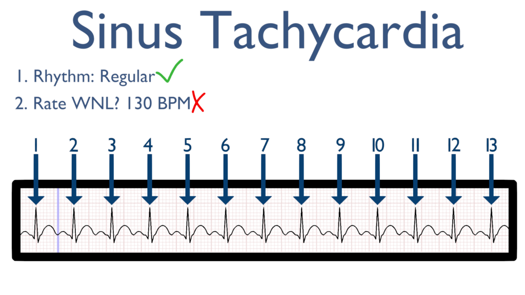
1. Assess the rhythm.
First, the rate should be regular. This can be identified by regularly spaced R waves.
2. Assess the rate.
Next, when assessing the rate you will find it is greater than 100 BPM.
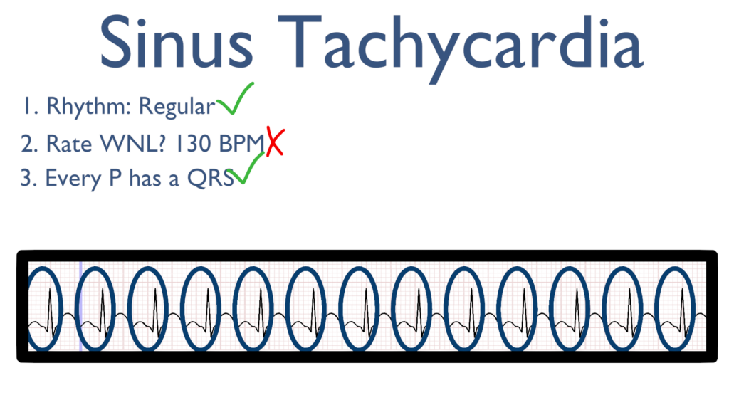
3. Assess atrial and ventricular rates.
Here we will see a QRS directly after every P wave.
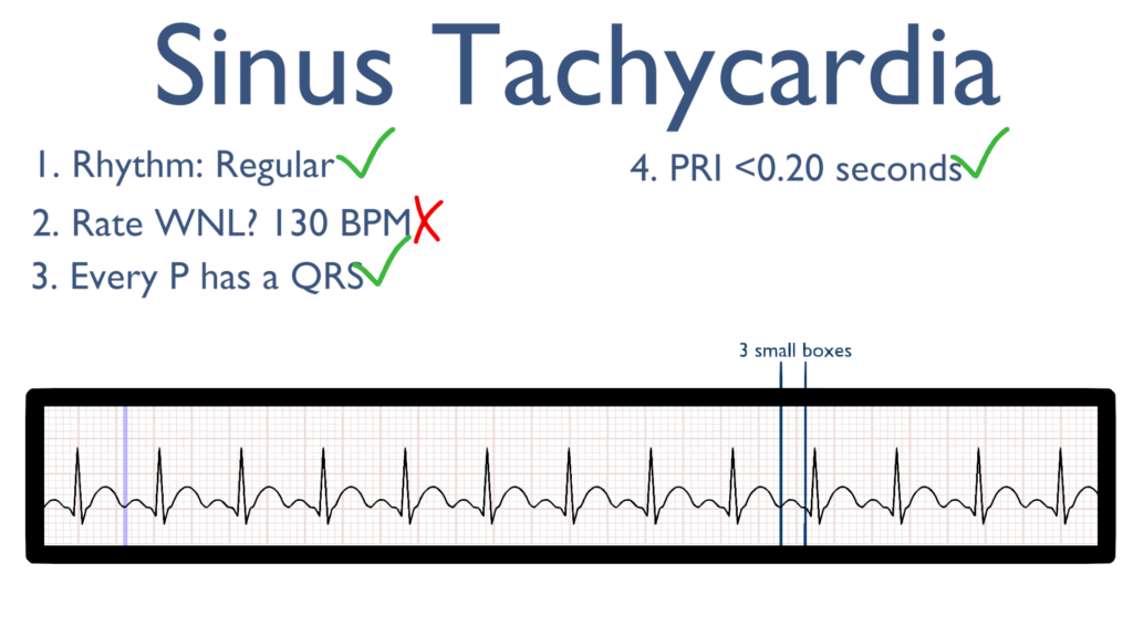
4. Assess the P waves and PR interval.
Now, take a look at the P waves and assess the PRI. All P waves should deflect in the positive direction (upwards).
The PRI should be within 0.12 sec to 0.20 sec.
Better yet, PRI should be between 3 to 5 small boxes.
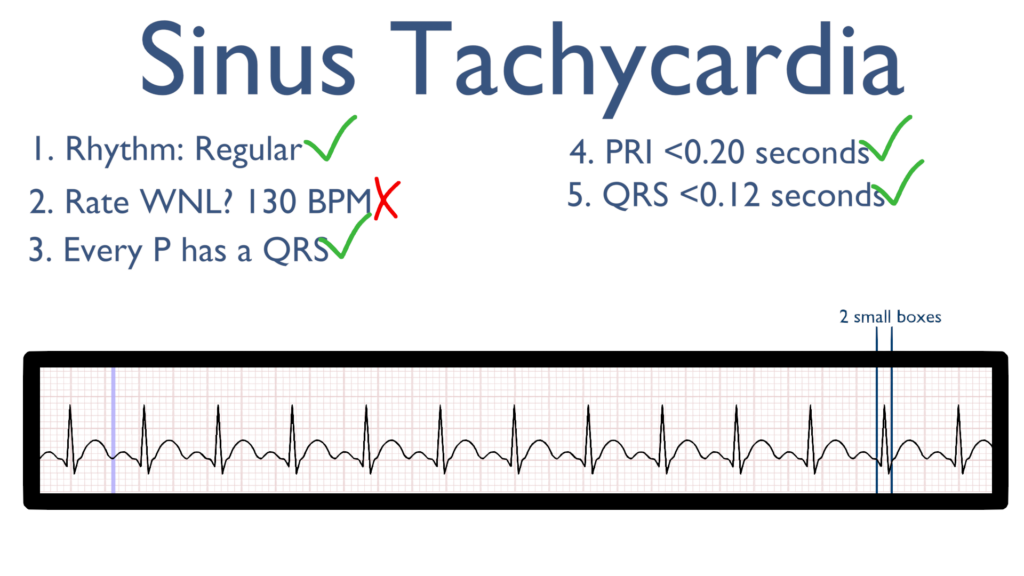
5. Assess the QRS.
Here take a look at the QRS complex.
The QRS should be between 0.06 seconds to 0.12 seconds.
Better yet, QRS should be between 1.5 to 3 small boxes.
6. Assess the T waves.
T waves should deflect in the positive direction with no ST-Elevation or ST-Depression.
The QT Interval should also be between 0.35 seconds to 0.45 seconds.
Easier to remember, the QT interval should be, roughly, between 9 to 12 small boxes.
7. Assess for Ectopy.
As you can see on this 6 second strip there are no ectopic beats. This is to say there are no PVC’s or PAC’s present.
Treatments for Sinus Tachycardia
In order to treat Sinus Tach it is important to understand the underlying cause. While tachycardia may be caused by an underlying cardiac disease it also may be caused by stress, exercise, pain, infection, hypovolemia, respiratory disorders, and abnormal electrolytes, etc.
Consider clinical presentation, lab work, and the H’s and T’s of the ACLS algorithms and then treat the underlying cause.
Fluids, antibiotics, antipyretics, and respiratory medications may be given to treat the underlying causes.
Cardiac medications may be given to slow the heart rate depending upon disease process.
Cardiac Medications: