So, in the previous article, ECG Impulse Basics, we covered electrical impulse flow through the heart. Here we will talk briefly about how we capture that flow. These concepts will be explored in depth within their own individual articles.
Electrocardiogram, or ECG and EKG for short, is a graphical representation of electrical flow through the heart. Monitoring someone’s ECG is a great way to understand how their heart is working because the electrical current is directly related to the heart’s muscular contractions which is directly related to blood flow throughout the body.
**On a side note ECG and EKG are used interchangeably. A Dutch scientist named Willem Einthoven first discovered how to visualize electrical conductivity through the heart. Remember his name. His original concept still plays a major role today in cardiac monitoring. The Dutch used the Germanic form of cardio, with a k, when naming it.This led it to be known as an EKG, or electrokardiogram. So, if you see EKG or ECG just know that these mean the same thing.**
Ok, so to start, let’s cover the different types of ECG setups. It is important to note that some setups are made to monitor cardiac electrical function and others are made to diagnose cardiac electrical function. We will identify which is which as we go through this module.
To begin, when we talk about ECG we are talking about how electrodes placed on the skin in specific areas of the body pick up and interpret electrical impulse. The basic set up originally created by Willem Einthoven was the 3 lead ECG. This 3 lead system is still used today. From there were begin to add electrodes to make up the 5 lead, the 6 lead, and the 12 lead systems.
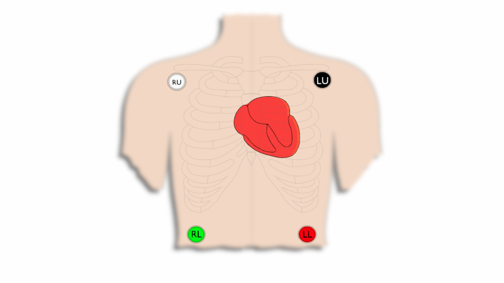
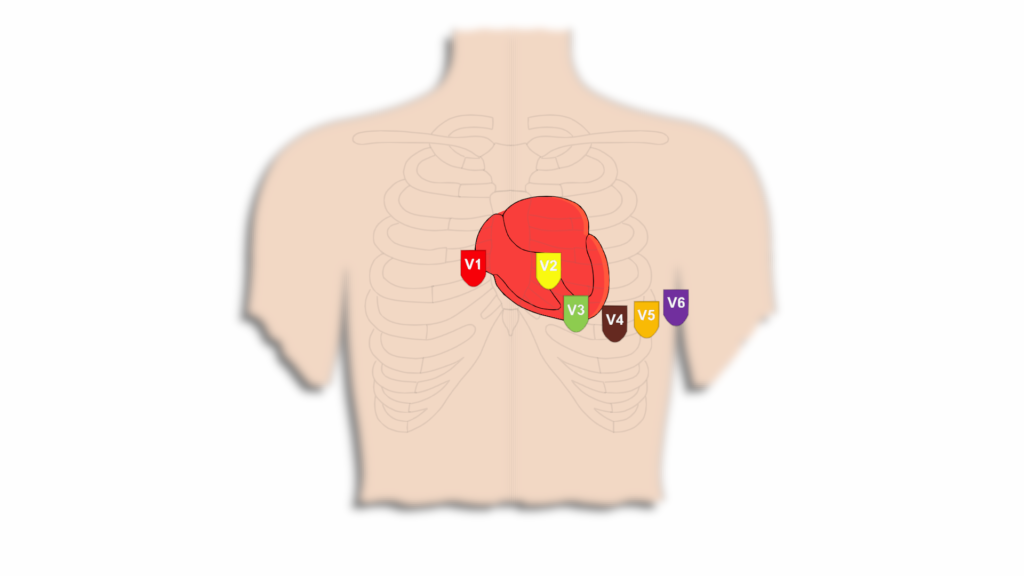
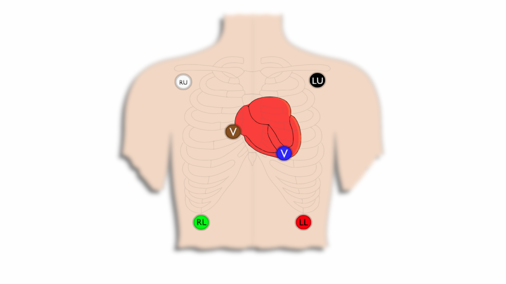
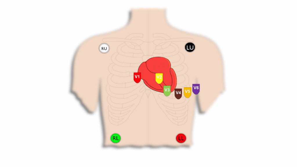
These electrodes can either be limb electrodes or precordial (in front of the heart) electrodes. Limbs leads consist of Right Upper/Arm, Left Upper/Arm, Right Lower/Leg, Left Lower/Leg. Precordial leads are the V leads and consist of V1, V2, V3, V4, V5, V6.
Limb Leads
- Set up on torso in 3, 5, 6 Lead set ups
- Set up on limbs for 12 Lead
Precordial Leads
- This shows a standard left side set up for precordial leads
- With 5 lead setup “V” is in the place of V1
- With 6 lead setup “Va” is in the place of V1 and “Vb” is in the place of V4
When it comes to these ECG setups the 3-lead, 5-lead, and 6-lead setups are used for cardiac monitoring. They are not meant to diagnose but to allow us to monitor cardiac function. The 12-lead set up is the gold standard that is utilized to diagnose cardiac function. We can still see and categorize dysrhythmias with the monitoring setups but to appropriately diagnose cardiac function we need to capture it on a 12-Lead.
Below we will show the basic set ups for each of these.
3 Lead ECG
- 3 Electrodes used
- Provides 6 different views of the heart
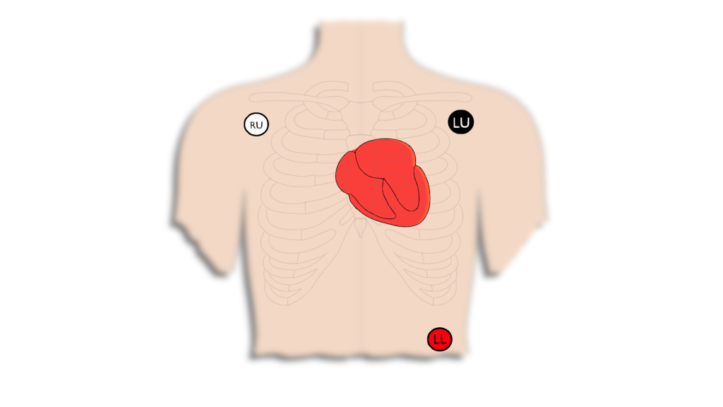
5 Lead ECG
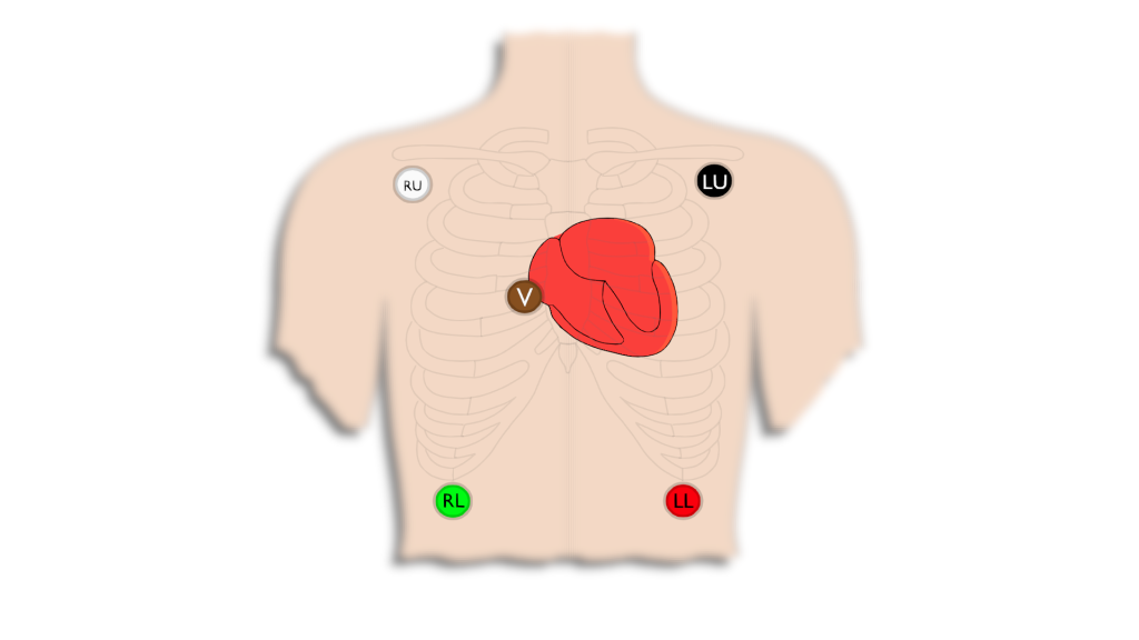
- 2 more electrodes added to the 3 lead system
- 7 total views
- 6 of the same views from the 3 Lead setup plus 1 additional view “V”
- The RL electrode does not provide a view but acts as a “grounding” lead
6 Lead ECG
- 1 more electrode added to the 5 Lead system
- “V” leads are labeled “Va” and “Vb”.
- 8 total views at a time
- 7 of the same views from the 6 Lead. plus 1 additional view from “Vb”
- “V” electrodes can be placed throughout the various 12 lead landmarks to show many different views.
12 Lead ECG
- Gold standard of diagnostics
- 12 total views made up from 10 electrodes
- Left sided is the most typical set up used
- Precordial leads can be set up either on right side or even on the patients back to diagnose different pathologies.
Read next: 3 Lead ECG